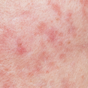
Post-inflammatory hyperpigmentation after carbon dioxide laser: review of prevention and risk factors

Accepted: 26 April 2023
APPENDIX 1: 64
HTML: 9
All claims expressed in this article are solely those of the authors and do not necessarily represent those of their affiliated organizations, or those of the publisher, the editors and the reviewers. Any product that may be evaluated in this article or claim that may be made by its manufacturer is not guaranteed or endorsed by the publisher.
The CO2 laser has been widely utilized in dermatology; its expanding clinical applications include the management of neoplastic lesions, benign growths, cosmetic conditions, and reactive disorders. The laser’s popularity is mainly due to the high precision and short recovery time this technology provides. However, postinflammatory hyperpigmentation (PIH) has been one of the challenging adverse effects of the CO2 laser. Therefore, several modalities have been studied for the prevention of PIH following CO2 laser treatment. This review aims to analyze the incidence of PIH after CO2 laser therapy, identify its risk factors, and assess the efficacy of the examined treatment modalities in preventing PIH. Pubmed and Embase databases were searched for this study, and relative clinical trials were included in the review. Descriptive findings – including age, gender, skin type, types of intervention, and incidence of PIH – were reported. When appropriate, the incidence of PIH was compared across each possible individual factor, such as skin type, gender, and type of intervention. A total of 211 articles were identified, and 14 relevant articles were included in this review. Seventy percent of the subjects were females (n=219), and 30% were males (n=94), with a mean age of 30 years (SD=7.8). The most common skin types were type IV (59%) followed by type III (25%). In total, eight studies investigated the prevention of PIH. The incidence of PIH after CO2 laser significantly varies between studies and differs based on the type of intervention. The studies indicate that the use of Clobetasol propionate 0.05% and fusidic acid cream appeared to effectively reduce PIH, recording an incidence rate of 39% and 53.3%, respectively. The Fitzpatrick-skinphenotype did not appear to influence the risk of PIH. There is a lack of high-powered clinical studies analyzing the incidence of PIH after CO2 laser treatment and the associated risk factors. PIH occurrence may be related to inflammation resulting from thermal damage by the CO2 laser. Consequently, the use of postoperative topical medications with anti-inflammatory properties might reduce its incidence. The use of ultra-potent topical corticosteroids and topical fusidic acid appeared to reduce PIH, possibly reducing postoperative inflammation effectively. Similarly, platelet-containing plasma may be beneficial in reducing CO2 side effects, including PIH. However, more studies are needed to further establish the influence of skin type on PIH and investigate modalities to reduce PIH occurrence after CO2 laser use.
Conforti C, Vezzoni R, Giuffrida R, et al. An overview on the role of CO2 laser in general dermatology. Dermatol Ther 2021;34:e14692.
Kaplan I. The CO2 surgical laser. Photomed Laser Surg 2010;28:847-8.
Cheyasak N, Manuskiatti W, Maneeprasopchoke P, Wanitphakdeedecha R. Topical corticosteroids minimise the risk of postinflammatory hyper-pigmentation after ablative fractional CO2 laser resurfacing in Asians. Acta Derm Venereol 2015;95:201-5.
Kim J, Kim B, Kim S, et al. The effect of human umbilical cord blood-derived mesenchymal stem cell media containing serum on recovery after laser treatment: a double-blinded, randomized, split-face controlled study. J Cosmet Dermatol 2020;19:651-6.
Moon HR, Yun WJ, Lee YJ, et al. A prospective, randomized, double-blind comparison of an ablative fractional 2940-nm erbium-doped yttrium aluminum garnet laser with a nonablative fractional 1550-nm erbium-doped glass laser for the treatment of photoaged Asian skin. J Dermatolog Treat 2015;26:551-7.
Rokhsar CK, Ciocon DH. Fractional photothermolysis for the treatment of postinflammatory hyperpigmentation after carbon dioxide laser resurfacing. Dermatol Surg 2009;35:535-7.
Wat H, Wu DC, Chan HH. Fractional resurfacing in the Asian patient: current state of the art. Lasers Surg Med 2017;49:45-59.
Kim JS, Ginter A, Ranjit-Reeves R, Woodward JA. Patient satisfaction and management of postoperative complications following ablative carbon dioxide laser resurfacing of the lower eyelids. Ophthalmic Plast Reconstr Surg 2021;37:450-6.
Sarnoff D, Gotkin H, Doerfler B, et al. The safety of laser skin resurfacing with the microablative carbon dioxide laser and review of the literature. J Drugs Dermatol 2018;17:1157-62.
Bernstein LJ, Kauvar AN, Grossman MC, Geronemus RG. The short- and long-term side effects of carbon dioxide laser resurfacing. Dermatol Surg 1997;23:519-25.
Gad SE, Neinaa YME, Rizk OK, Ghaly NER. Efficacy of platelet-poor plasma gel in combination with fractional CO(2) laser in striae distensae: a clinical, histological, and immunohistochemical study. J Cosmet Dermatol 2021;20:3236-44.
Goh C. Management of post-acne scars in Asians-need for a paradigm shift? In: Australasian Journal of Dermatology. Wiley: Hoboken NJ USA; 2017. pp 57-57.
Jimenez JC, Montes JR, Maldonado J. Aesthetic benefits of CO2 laser photorejuvenation treatment for malar mounds (festoons). Invest Ophthalmol Visual Sci 2015;56:4735.
Wei M, Li L, Zhang XF, et al. Fusidic acid cream comparatively minimizes signs of inflammation and postinflammatory hyperpigmentation after ablative fractional CO2 laser resurfacing in Chinese patients: a randomized controlled trial. J Cosmet Dermatol 2021;20:1692-9.
Techapichetvanich T, Wanitphakdeedecha R, Iamphonrat T, et al. The effects of recombinant human epidermal growth factor containing ointment on wound healing and post inflammatory hyperpigmentation prevention after fractional ablative skin resurfacing: a split-face randomized controlled study. J Cosmet Dermatol 2018;17:756-61.
Lueangarun S, Srituravanit A, Tempark T. Efficacy and safety of moisturizer containing 5% panthenol, madecassoside, and copper-zinc-manganese versus 0.02% triamcinolone acetonide cream in decreasing adverse reaction and downtime after ablative fractional carbon dioxide laser resurfacing: A split-face, double-blinded, randomized, controlled trial. J Cosmet Dermatol 2019;18:1751-7.
Neinaa YME, Gheida SF, Mohamed DAE. Synergistic effect of platelet-rich plasma in combination with fractional carbon dioxide laser versus its combination with pulsed dye laser in striae distensae: a comparative study. Photodermatol Photoimmunol Photomed 2021;37:214-23.
Shin S, Shin JU, Lee Y, et al. The effects of a multigrowth factor-containing cream on recovery after laser treatment: a double-blinded, randomized, split-face controlled study. J Cosmet Dermatol 2017;16:76-83.
Lueangarun S, Tempark T. Efficacy of MAS063DP lotion vs 0.02% triamcinolone acetonide lotion in improving post-ablative fractional CO2 laser resurfacing wound healing: a split-face, triple-blinded, randomized, controlled trial. Int J Dermatol 2018;57:480-7.
Alster TS, Nanni CA, Williams CM. Comparison of four carbon dioxide resurfacing lasers. A clinical and histopathologic evaluation. Dermatol Surg 1999;25:153-9.
Al Mohizea S. The effect of menstrual cycle on laser induced hyperpigmentation. J Drugs Dermatol 2013;12:1335-6.
Tan KL, Kurniawati C, Gold MH. Low risk of postinflammatory hyperpigmentation in skin types 4 and 5 after treatment with fractional CO2 laser device. J Drugs Dermatol 2008;7:774-7.
Elmorsy EH, Elgarem YF, Sallam ES, Taha AAA. Fractional carbon dioxide laser versus carboxytherapy in treatment of striae distensae. Lasers Surg Med 2021;53:1173-9.
Suh JH, Lee SK, et al. Efficacy of bleomycin application on periungual warts after treatment with ablative carbon dioxide fractional laser: a pilot study. J Dermatolog Treat 2020;31:410-4.
Al-Muriesh M, Huang CZ, Ye Z, Yang J. Dermoscopy and VISIA imager evaluations of non-insulated microneedle radiofrequency versus fractional CO2 laser treatments of striae distensae. J Eur Acad Dermatol Venereol 2020;34:1859-66.
Wu PP, He H, Hong WD, et al. The biological evaluation of fusidic acid and its hydrogenation derivative as antimicrobial and anti-inflammatory agents. Infect Drug Resist 2018;11:1945-57.
Grimes PE. Management of hyperpigmentation in darker racial ethnic groups. Semin Cutan Med Surg 2009;28:77-85.
Ruiz-Maldonado R, Orozco-Covarrubias ML. Postinflammatory hypopigmentation and hyperpigmentation. Semin Cutan Med Surg 1997;16:36-43.
Sriprachya-anunt S, Marchell NL, Fitzpatrick RE, et al. Facial resurfacing in patients with Fitzpatrick skin type IV. Lasers Surg Med 2002;30:86-92.
West TB, Alster TS. Effect of pretreatment on the incidence of hyperpigmentation following cutaneous CO2 laser resurfacing. Dermatol Surg 1999;25:15-17.
Takiwaki H, Shirai S, Kohno H, et al. The degrees of UVB-induced erythema and pigmentation correlate linearly and are reduced in a parallel manner by topical anti-inflammatory agents. J Invest Dermatol 1994;103:642-6.
Singh PK, Singh G. Relative potency of topical corticosteroid preparations. Indian J Dermatol Venereol Leprol 1985;51:309-12.
Uaboonkul T, Nakakes A, Ayuthaya PK. A randomized control study of the prevention of hyperpigmentation post Q-switched Nd:YAG laser treatment of Hori nevus using topical fucidic acid plus betamethasone valerate cream versus fucidic acid cream. J Cosmet Laser Ther 2012;14:145-9.
Doghaim NN, El‐Tatawy RA, Neinaa YMEH. Assessment of the efficacy and safety of platelet poor plasma gel as autologous dermal filler for facial rejuvenation. J Cosmet Dermatol 2019;18:1271-9.
Demidova-Rice TN, Hamblin MR, Herman IM. Acute and impaired wound healing: pathophysiology and current methods for drug delivery, part 2: role of growth factors in normal and pathological wound healing: therapeutic potential and methods of delivery. Adv Skin Wound Care 2012;25:349.
Saitta P, Krishnamurthy K, Brown LH. Bleomycin in dermatology: a review of intralesional applications. Dermatol Surg 2008;34:1299-313.
Puder JJ, Blum CA, Mueller B, et al. Menstrual cycle symptoms are associated with changes in low-grade inflammation. Eur J Clin Invest 2006;36:58-64.
Copyright (c) 2023 the Author(s)

This work is licensed under a Creative Commons Attribution-NonCommercial 4.0 International License.
PAGEPress has chosen to apply the Creative Commons Attribution NonCommercial 4.0 International License (CC BY-NC 4.0) to all manuscripts to be published.


 https://doi.org/10.4081/dr.2023.9703


