Pigmentary mammary Paget disease: Clinical, dermoscopical and histological challenge

HTML: 14
All claims expressed in this article are solely those of the authors and do not necessarily represent those of their affiliated organizations, or those of the publisher, the editors and the reviewers. Any product that may be evaluated in this article or claim that may be made by its manufacturer is not guaranteed or endorsed by the publisher.
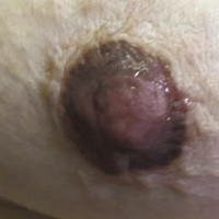
A very rare variant of mammary Paget disease (MPD) is the pigmented MPD, first described in 1956. It is very difficult to distinguish this variant from melanoma both clinically and dermoscopically. The diagnosis is confirmed by histopathology and immunohistochemistry. Correct diagnosis is crucial for surgical treatment, which is different for these two diseases. We report the case of a 92-year-old woman, who presented an asymptomatic pigmented lesion of the right nipple and areola. The lesion was arisen for about 6 months and was suspected for melanoma because of clinical and dersmoscopic characteristics. Incisional biopsy revealed tumor cells, that proliferate in the major mammary ducts, and tumor cells in the overlying epidermis of the nipple, thus diagnosing pigmented mammary Paget disease. The patient underwent radical mastectomy.
PAGEPress has chosen to apply the Creative Commons Attribution NonCommercial 4.0 International License (CC BY-NC 4.0) to all manuscripts to be published.


 https://doi.org/10.4081/dr.2021.9235


