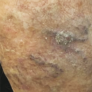
Granular cell dermatofibroma: a potential diagnostic pitfall

Accepted: 13 October 2021
HTML: 113
All claims expressed in this article are solely those of the authors and do not necessarily represent those of their affiliated organizations, or those of the publisher, the editors and the reviewers. Any product that may be evaluated in this article or claim that may be made by its manufacturer is not guaranteed or endorsed by the publisher.
Dermatofibroma, also known as fibrous histiocytoma, is one of the most common cutaneous soft-tissue tumours. Many variants of dermatofibromas have been described and knowledge of these variations is important to avoid a misdiagnosis of a possibly more aggressive tumor. Histological features of different variants can coexist in the same lesion, but typical common fibrous histiocytoma features are generally found, at least focally, in all cases. However, when cellular changes make up the majority of the lesion, the histopathological diagnosis can become more complex and requires immunohistochemical investigations for a correct nosographic classification. We report on the case of a cutaneous fibrous histiocytoma, “granular cell” variant, found on the left leg of a 74-year old woman.
David J. Myers, Eric P. Fillman. Dermatofibroma. StatPearls Publishing 2020.
Han TY, Chang HS, Lee JH, et al. A clinical and histopathological study of 122 cases of dermatofibroma (benign fibrous histiocytoma). Ann Dermatology 2011;23:185-92. DOI: https://doi.org/10.5021/ad.2011.23.2.185
LeBoit PE, Barr RJ, Bural S, et al. Primitive polypoid granular-cell tumor and other granular-cell neoplasms of apparent not neural origin. Am J Surg Pathol 1991;15:48–58. DOI: https://doi.org/10.1097/00000478-199101000-00006
Aloi F, Albertazzi D, Pippione M. Dermatofibroma with Granular Cells: A Report of Two Cases. Department of Dermatology, University of Turin, Italy. Dermatology 1999;199:54–56. DOI: https://doi.org/10.1159/000018179
Lee J. Epithelioid Cell Histiocytoma With Granular Cells. (Another Nonneural Granular Cell Neoplasm). Am J Dermatopathology 2007;29:475–6. DOI: https://doi.org/10.1097/DAD.0b013e31813735a8
Perret RE, Jullie ML, Vergier B, et al. A subset of so-called dermal non-neural granular cell tumours are underlined by ALK fusions, further supporting the idea that they represent a variant of epithelioid fibrous histiocytoma. Histopathology 2018;3:532-4. DOI: https://doi.org/10.1111/his.13645
Soukup J, Hadzi-Nikolov D, Pol RA. Dermatofibroma-like granular cell tumour: a potential diagnostic pitfall. J Pathol 2016;3:291-4. DOI: https://doi.org/10.5114/pjp.2016.63782
Alves JV, Matos DM, Barreiros HF, Bártolo EA. Variants of dermatofibroma--a histopathological study. Ann Bras Dermatol 2014;3:472-7. DOI: https://doi.org/10.1590/abd1806-4841.20142629
Cheng SD, Usmani AS, DeYoung BR, et al. Dermatofibroma-like Granular Cell Tumor. J Cutan Pathol 2001;1:49-52. DOI: https://doi.org/10.1034/j.1600-0560.2001.280106.x
Yeshwant BR, Dodson TB. S-100 Negative Granular Cell Tumor (So-called Primitive Polypoid Non-neural Granular Cell Tumor) of the Oral Cavity. Case Reports Head Neck Pathol 2017;3:404-12. DOI: https://doi.org/10.1007/s12105-016-0760-3
Martin Y, Sathyakumar M, Premkumar J, Magesh KT. Granular Cell Ameloblastoma; Case Reports. J Oral Maxillofac Pathol 2017;21:183. DOI: https://doi.org/10.4103/jomfp.JOMFP_45_15
Banerjee SS, Harris M, Eyden BP, Hamid BN. Granular cell variant of dermatofibrosarcoma protuberans. Histopathology 1990;4:375-8. DOI: https://doi.org/10.1111/j.1365-2559.1990.tb00745.x
Cardis MA, Ni J, Bhawan J. Granular cell differentiation: A review of the published work. J Dermatol 2017;44:251-8. DOI: https://doi.org/10.1111/1346-8138.13758
Copyright (c) 2022 the Author(s)

This work is licensed under a Creative Commons Attribution-NonCommercial 4.0 International License.
PAGEPress has chosen to apply the Creative Commons Attribution NonCommercial 4.0 International License (CC BY-NC 4.0) to all manuscripts to be published.


 https://doi.org/10.4081/dr.2022.9110


