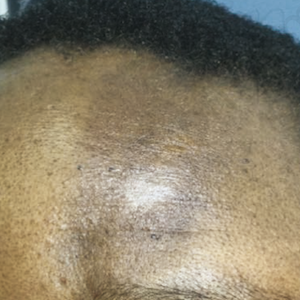
En coup de sabre morphea: an uncommon condition in Africa
HTML: 38
All claims expressed in this article are solely those of the authors and do not necessarily represent those of their affiliated organizations, or those of the publisher, the editors and the reviewers. Any product that may be evaluated in this article or claim that may be made by its manufacturer is not guaranteed or endorsed by the publisher.
The term en coup de sabre morphea refers to a lesion of linear morphea typically located in the frontoparietal scalp and/or the paramedian forehead, often resembling a strike with a sword. In literature, en coup de sabre morphea, and en coup de sabre scleroderma are terms used interchangeably and synonymously. Due to the rarity of this condition, treatment is largely based on case report series, leaving much room for speculation in terms of drugs of choice, duration of treatment, and dosages. Although it typically leaves behind notable and often permanent skin pigmentary changes and indentation of the affected areas, this condition usually remits spontaneously, even in the absence of an active form of treatment. The disease severity and prognosis vary according to the subtype: circumscribed morphea has a generally more benign course when compared with linear scleroderma and generalized morphea.
Holland KE, Steffes B, Nocton JJ, et al. Linear scleroderma en coup de sabre with associated neurologic abnormalities. Pediatrics 2006;117:e132-6.
Albuquerque JVD, Andriolo BN, Vasconcellos MR, et al. Interventions for morphea. Cochrane Database Syst Rev 2019;7:CD005027.
Megha M, Witman PM. En coup de sabre morphea and parry-romberg syndrome: a retrospective review of 54 patients. J Am Acad Dermatol 2007;56:257-63.
Penmetsa GK, Sapra A. Morphea. Treasure Island (FL), USA: StatPearls Publishing; 2023.
O’Brien JC, Nymeyer H, Green A, Jacobe HT. Changes in disease activity and damage over time in patients with morphea. JAMA Dermatol 2020;156:513-20.
Khatri S, Torok KS, Mirizio E, et al. Autoantibodies in morphea: an update. Front Immunol 2019;10:1487.
Zulian F, Athreya BH, Laxer R, et al. Juvenile localized scleroderma: clinical and epidemiological features in 750 children. An international study. Rheumatology 2006;45:614-20.
Saceda-Corralo D, Tosti A. Trichoscopic features of linear morphea on the scalp. Skin Appendage Disord 2018;4:31-3.
Fledelius CH, Danielsen PL, Ullman S. Ophthalmic findings in linear scleroderma manifesting as facial en coup de sabre. Eye 2018;32:1688-96.
Dixit S, Kalkur C, Sattur AP, et al. Scleroderma and dentistry: two case reports. J Med Case Rep 2016;10:297.
Lauesen SR, Daugaard Jensen J, Lauridsen EF, Kjær I, Localised scleroderma en coup de sabre affecting the skin, dentition and bone tissue within craniofacial neural crest fields. Clinical and radiographic study of six patients. Eur Arch Paediatr Dent 2019;20:339-50.
Taniguchi T, Asano Y, Tamaki Z, et al. Histological features of localized scleroderma ‘en coup de sabre’: a study of 16 cases. J Eur Acad Dermatol Venereol 2014;28:1805-10.
Pedowska M, Ptasiewicz M, Szumiło J, Chałas R. Morphological and clinical picture of the morphea in the oral cavity. Folia Morphol 2022;81:544-50.
Chiu YE, Vora S, Kwon EKM, Maheshwari M. A significant proportion of children with morphea en coup de sabre and parry-romberg syndrome have neuroimaging findings. Pediatr Dermatol 2012;29:738-48.
Graham PM, Gupta N, Altman DA. En coup de sabre. Cutis 2019;103:34-6.
Copyright (c) 2022 the Author(s)
This work is licensed under a Creative Commons Attribution-NonCommercial 4.0 International License.
PAGEPress has chosen to apply the Creative Commons Attribution NonCommercial 4.0 International License (CC BY-NC 4.0) to all manuscripts to be published.