Wickham striae on skin appendages: a helpful dermoscopic feature


Published: 6 June 2023
Abstract Views: 745
PDF: 272
HTML: 7
Publisher's note
All claims expressed in this article are solely those of the authors and do not necessarily represent those of their affiliated organizations, or those of the publisher, the editors and the reviewers. Any product that may be evaluated in this article or claim that may be made by its manufacturer is not guaranteed or endorsed by the publisher.

Authors

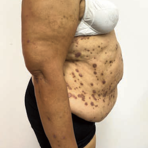
Lichen planus (LP) is a chronic inflammatory disease, clinically characterized by purpuric, itchy papules that typically spread on the trunk and extremities. Other body sites can also be affected, including mucosal membranes, nails, and the scalp. The use of dermoscopy is essential in determining the diagnosis of LP, as it may highlight pathognomonic features such as Wickham striae (WS). WS are thin, pearly white structures arranged in a reticular pattern that is observed over LP lesions and histologically correspond to epidermal hypergranulosis. WS is usually most visible on the oral mucosa but can also cover almost every active LP papule. Here, we report two cases of biopsy-proven LP that show WS on dermoscopy in two specific sites: the scalp and proximal nail fold.


Boyd AS, Neldner KH. Lichen planus. J Am Acad Dermatol 1991;25:593-619. DOI: https://doi.org/10.1016/0190-9622(91)70241-S

Usatine RP, Tinitigan M. Diagnosis and treatment of lichen planus. Am Fam Physician 2011;84:53-60.

Alrashdan MS, Cirillo N, McCullough M. Oral lichen planus: a literature review and update. Arch Dermatol Res 2016;308:539-51. DOI: https://doi.org/10.1007/s00403-016-1667-2

Lallas A, Zalaudek I, Argenziano G, et al. Dermoscopy in general dermatology. Dermatol Clin 2013;31:679-94. DOI: https://doi.org/10.1016/j.det.2013.06.008

Lallas A, Kyrgidis A, Tzellos TG, et al. Accuracy of dermoscopic criteria for the diagnosis of psoriasis, dermatitis, lichen planus and pityriasis rosea. Br J Dermatol 2012;166:1198-1205. DOI: https://doi.org/10.1111/j.1365-2133.2012.10868.x

Atzmony L, Reiter O, Hodak E,et al. Treatments for Cutaneous Lichen Planus: A Systematic Review and Meta-Analysis. Am J Clin Dermatol 2016;17:11-22. DOI: https://doi.org/10.1007/s40257-015-0160-6

Iorizzo M, Tosti A, Starace M, et al. Isolated nail lichen planus: An expert consensus on treatment of the classical form. J Am Acad Dermatol 2020;83:1717-23 DOI: https://doi.org/10.1016/j.jaad.2020.02.056

Steffen C, Dupree ML. Louis-Frédéric Wickham and the Wickham's striae of lichen planus. Skinmed 2004;3:287-9. DOI: https://doi.org/10.1111/j.1540-9740.2004.02647.x

Sachdeva S, Sachdeva S, Kapoor P. Wickham striae: etiopathogenensis and clinical significance. Indian J Dermatol 2011;56:442-3. DOI: https://doi.org/10.4103/0019-5154.84739

Rouai M, Litaiem N, Hammami H, et al. Dermoscopic features of mucosal lichen planus. Int J Dermatol 2021;60:1368-72. DOI: https://doi.org/10.1111/ijd.15683

. Rivers JK, Jackson R, Orizaga M. Who was Wickham and what are his striae? Int J Dermatol 1986;25:611–3 DOI: https://doi.org/10.1111/j.1365-4362.1986.tb04716.x

Summerly R, Wilson Jones E. The Microarchitecture of Wickham's Stirae. Trans St. Jhon's Hosp Dermatol Soc. 1964;50:157–61.

Ryan TJ. The direction of the growth of the epithelium. Br J Dermatol 1966;78:403–15. DOI: https://doi.org/10.1111/j.1365-2133.1966.tb12237.x

Roja Renuka S, Manoharan K. Dermoscopy-a simple diagnostic and prognostic tool in lichen planus. QJM 2022;115:109-10. DOI: https://doi.org/10.1093/qjmed/hcab318

Papageorgiou C, Apalla Z, Lazaridou E, et al . Atypical case of lichen planus recognized by dermoscopy. Dermatol Pract Concept 2016;6:39-42. DOI: https://doi.org/10.5826/dpc.0604a09

Fagotto, L., Gnesotto, L., Vincenzi, C., Piraccini, B. M., Naldi, L., & Sechi, A. (2023). Wickham striae on skin appendages: a helpful dermoscopic feature. Dermatology Reports, 15(3). https://doi.org/10.4081/dr.2023.9698

Downloads

Download data is not yet available.

Citations